Ethan Bendau
Mentors: Distinguished Professor Robert Alfano and Dr. Lingyan Shi, Institute for Ultrafast Spectroscopy and Lasers, CCNY
Fundamental concepts of physics are at the heart of virtually every aspect of medical imaging. PET scans detect matter-antimatter annihilation events, MRI machines manipulate nuclear spin states, and x-ray images show differential absorption of high energy electromagnetic radiation by different types of tissue. These techniques are critical for breast cancer detection, but detailed information on molecular characteristics, which foretell various risks, requires invasive biopsy. The salient properties of visible light show promise in further understanding and detecting disease. Triple-negative breast cancer (TNBC) is an aggressive breast cancer subtype that lacks three hormone receptors used to detect and treat other types of cancer. It also disproportionately affects black and Hispanic women. TNBC carries a poor prognosis and early detection is crucial for improved patient outcomes.
I will discuss our use of multiphoton fluorescence microscopy (MPM) and resonance Raman spectroscopy (RRS) as a rapid, label-free means of TNBC cancer diagnosis in mice by detecting intrinsic protein fluorescence and unique spectral fingerprints, respectively, that are associated with different tissue structures and metabolic activity.
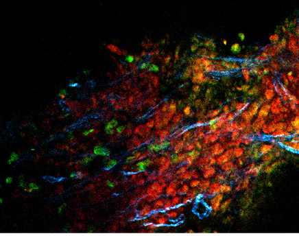
Multiphoton microscope image showing fluorescence from NADH (green) and FAD (red) as well as second harmonic generation from collagen (blue) in a mouse triple-negative breast cancer tumor.
Last Updated: 04/27/2018 12:24